Pre-implantation genetic diagnosis (PGD) is a laboratory procedure used in conjunction with in vitro fertilization (IVF) to reduce the risk of passing on inherited conditions. Some of the most common reasons for PGD are specific single-gene conditions (such as cystic fibrosis or sickle cell anemia) and structural changes of a parent's chromosomes. Families may also use PGD when a member of the family needs a bone marrow donor, as a way to have a child who can provide matching stem cells.
Pre-Implantation Genetic Diagnosis
(415) 353-7397
Typically, couples in need of these techniques are not infertile but have a family history of a condition and want to reduce the risk of having another child with significant health issues or early death. Through generally available genetic screening, however, occasionally couples who are seeking fertility treatment are found to be at risk of passing on an inherited condition, and PGD may be an option for them.
PGD is available for almost any inherited condition for which the exact mutation is known. A unique test must be developed for each couple, however. This test design may take up to several months to complete before beginning an IVF cycle.
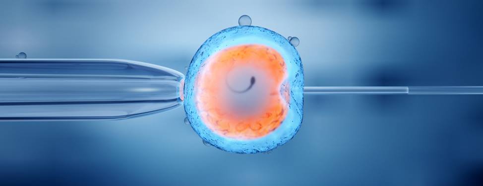
PGD uses IVF, in which multiple eggs are matured and retrieved. The oocytes — or primitive egg cells — are inseminated with a single sperm using intracytoplasmic sperm injection.
The resulting embryos are grown in culture until the six-to-eight-cell stage, which is day three of embryo development. At this point, the embryo is biopsied with the removal of one to two cells. This process does not damage the cells remaining within the embryo.
The isolated cells are evaluated for specific genetic conditions. Embryos that are found to be unaffected are transferred back to the woman's uterus on day five of embryo development.
Two main techniques are used for the genetic assessment:
- Polymerase chain reaction (PCR)In PCR, multiple copies of the gene of interest are made by a process of amplification. This amplification process allows the identification of very small amounts of DNA to make the diagnosis.
- Fluorescent in situ hybridization (FISH)FISH allows the laboratory to count the number of chromosomes in an isolated cell. This technique is used primarily for expected abnormalities in chromosome number, such as Down syndrome, or translocations (defects in the structure of the chromosome).
At UCSF, our embryology laboratory staff has extensive experience with embryo micromanipulation and biopsy. Our genetic counselor is available to coordinate your cycle with the IVF team and the PGD laboratory, to make the process as smooth as possible.
UCSF Health medical specialists have reviewed this information. It is for educational purposes only and is not intended to replace the advice of your doctor or other health care provider. We encourage you to discuss any questions or concerns you may have with your provider.


