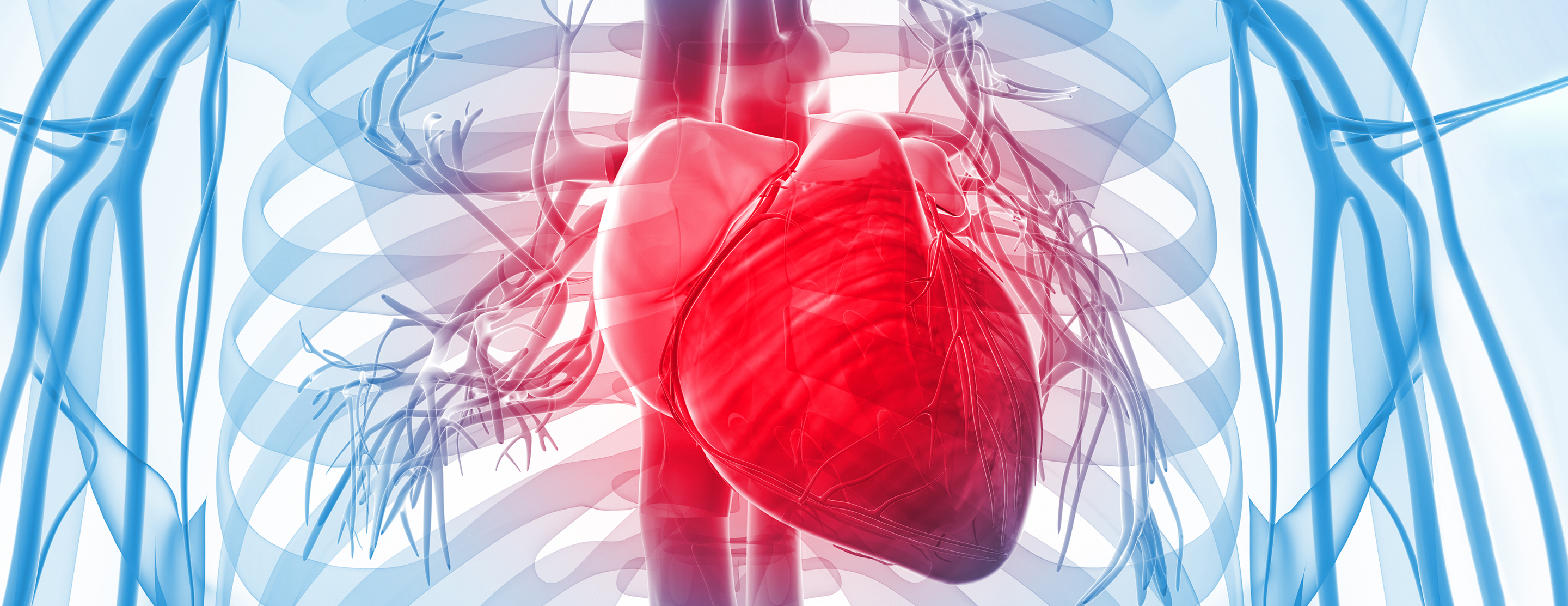
Alcohol septal ablation is a minimally invasive treatment for hypertrophic cardiomyopathy (HCM), an inherited disease in which parts of the heart muscle become abnormally thick, making it harder for the heart to pump blood through the body. In this procedure, alcohol is injected into the thickened area, causing some of the cells to shrink and die. Thinning the tissue allows blood to move through the heart more freely, alleviating many HCM symptoms.

Alcohol Septal Ablation
The procedure is less invasive than open-heart surgery, and patients recover much more quickly. We use a rigorous selection process to determine who is a good candidate for alcohol septal ablation, and the result is that most patients who receive this treatment gain relief from their symptoms.
Symptoms
In HCM, the abnormally thickened area is usually the ventricular septum, the muscular wall separating the lower right and left chambers (ventricles) of the heart. Many people with HCM have no symptoms. But if the septum bulges into the left ventricle and partially blocks blood flow out to the body, the heart has to work harder to pump; this can result in a variety of symptoms, including fatigue, shortness of breath and chest pain.
Evaluation
Patients receive a comprehensive evaluation at the UCSF Hypertrophic Cardiomyopathy Clinic. If you are a potential candidate for alcohol septal ablation, you will be seen by an interventional cardiologist, a heart specialist who performs these procedures. To get a better picture of your heart's condition, you may undergo the following tests prior to the procedure:
- Chest X-rays, to obtain images of the heart
- Blood tests
- Echocardiogram, an ultrasound of the heart, used to determine where the tissue is thickened
- Electrocardiogram (EKG), a recording of the heart’s electrical activity, which can provide information on the extent of damage and the heart chambers' size and position
Procedure
The procedure generally takes about two hours and requires general anesthesia, meaning that you will be completely asleep. Your doctor will determine the best approach for managing pain before, during and after the procedure.
For this minimally invasive procedure, the doctor makes small incisions in your neck and groin to reach arteries in those locations. A temporary pacemaker – to deliver electrical signals to your heart, if needed – is inserted into the artery in your neck. A catheter (small, flexible tube) is inserted into the artery in your groin and threaded through blood vessels to reach your heart. Your doctor will use advanced imaging to guide the catheter to the right place. Once there, a small amount of pure alcohol is slowly injected through the catheter into an artery that leads to the septum. The alcohol destroys part of the septum muscle. The catheter is removed from your body when the procedure is complete.
Recovery
After the procedure, we monitor you in a recovery area, where you'll spend several hours lying flat to prevent bleeding. You're then moved to our cardiac intensive care unit for two nights. Because a small percentage of patients develop heart block (a slowing heart rate) after this procedure, the temporary pacemaker is kept in your heart for at least two days. If you develop heart block and it persists, you may need to have a permanent pacemaker implanted. If no complications arise, you'll likely go home three days after the procedure.
It's important, after you leave the hospital, to follow all the instructions we give you regarding medications, exercise and diet, and care of the incision sites. Also, be sure to keep your follow-up appointments.
UCSF Health medical specialists have reviewed this information. It is for educational purposes only and is not intended to replace the advice of your doctor or other health care provider. We encourage you to discuss any questions or concerns you may have with your provider.


