


Bone scan
Definition
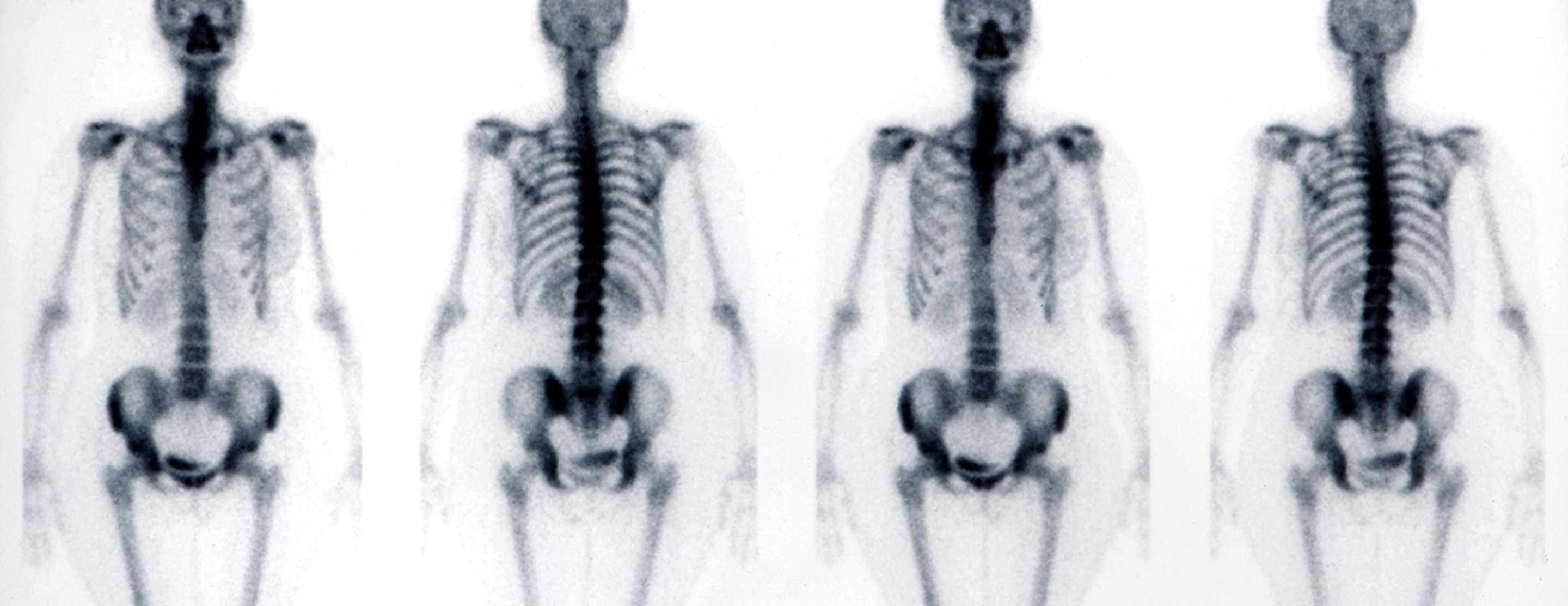
A bone scan is an imaging test used to diagnose bone diseases and find out how severe they are.
Alternative Names
Scintigraphy - bone
How the Test is Performed
A bone scan involves injecting a very small amount of radioactive material (radiotracer) into a vein. The substance travels through your blood to the bones and organs. As it wears off, it gives off a bit of radiation. This radiation is detected by a camera that slowly scans your body. The camera takes pictures of how much radiotracer collects in the bones.
If a bone scan is done to see if you have a bone infection, images may be taken shortly after the radioactive material is injected and again 3 to 4 hours later, when it has collected in the bones. This process is called a 3-phase bone scan.
To evaluate
The scanning part of the test will last about 1 hour. The scanner's camera may move above and around you. You may need to change positions.
You will probably be asked to drink extra water after you receive the radiotracer to keep the material from collecting in your bladder.
How to Prepare for the Test
You must remove jewelry and other metal objects. You may be asked to wear a hospital gown.
Tell your health care provider if you are or may be pregnant.
DO NOT take any medicine with bismuth in it, such as Pepto-Bismol, for 4 days before the test.
Follow any other instructions you are given.
How the Test will Feel
There is a small amount of pain when the needle is inserted. During the scan, there is no pain. You must remain still during the scan. The technologist will tell you when to change positions.
You may experience some discomfort due to lying still for a long period.
Why the Test is Performed
A bone scan is used to:
- Diagnose a bone tumor or cancer.
- Determine if a cancer that began elsewhere in your body has spread to the bones. Common cancers that spread to the bones include breast, lung, prostate, thyroid, and kidney.
- Diagnose a fracture, when it cannot be seen on a regular x-ray (most commonly hip fractures, stress fractures in the feet or legs, or spine fractures).
- Diagnose a
bone infection (osteomyelitis) . - Diagnose or determine the cause of bone pain, when no other cause has been identified.
- Evaluate metabolic disorders, such as
osteomalacia ,primary hyperparathyroidism ,osteoporosis ,complex regional pain syndrome , andPaget disease .
Normal Results
Test results are considered normal if the radiotracer is present evenly throughout all the bones.
What Abnormal Results Mean
An abnormal scan will show "hot spots" and/or "cold spots" as compared to surrounding bone. Hot spots are areas where there is an increased collection of the radioactive material. Cold spots are areas that have taken up less of the radioactive material.
Bone scan findings must be compared with other imaging studies, in addition to clinical information. Your provider will discuss any abnormal findings with you.
Risks
If you are pregnant or nursing, the test may be postponed to prevent exposing the baby to radiation. If you must have the test while breastfeeding, you should pump and throw away the breast milk for the next 2 days.
The amount of radiation injected into your vein is very small. All radiation is gone from the body within 2 to 3 days. The radiotracer that is used exposes you to a very small amount of radiation. The risk is probably no greater than with routine x-rays.
Risks related to the bone radiotracer are rare, but may include:
Anaphylaxis (severe allergic response)Rash Swelling
There is a slight risk of infection or bleeding when the needle is inserted into a vein.
References
Bearcroft PWP, Hopper MA. Imaging techniques and fundamental observations for the musculoskeletal system. In: Adam A, Dixon AK, Gillard JH, Schaefer-Prokop CM, eds. Grainger & Allison's Diagnostic Radiology: A Textbook of Medical Imaging. 6th ed. Philadelphia, PA: Elsevier Churchill Livingstone; 2015:chap 45.
Chernecky CC, Berger BJ. Bone scan (bone scintigraphy) - diagnostic. In: Chernecky CC, Berger BJ, eds. Laboratory Tests and Diagnostic Procedures. 6th ed. St Louis, MO: Elsevier Saunders; 2013:246-247.
Ribbens C, Namur G. Bone scintigraphy and positron emission tomography. In: Hochberg MC, Gravallese EM, Silman AJ, Smolen JS, Weinblatt ME, Weisman MH, eds. Rheumatology. 7th ed. Philadelphia, PA: Elsevier; 2019:chap 49.
Review Date: 02/24/2018
The information provided herein should not be used during any medical emergency or for the diagnosis or treatment of any medical condition. A licensed physician should be consulted for diagnosis and treatment of any and all medical conditions. Call 911 for all medical emergencies. Links to other sites are provided for information only -- they do not constitute endorsements of those other sites. Copyright ©2019 A.D.A.M., Inc., as modified by University of California San Francisco. Any duplication or distribution of the information contained herein is strictly prohibited.
Information developed by A.D.A.M., Inc. regarding tests and test results may not directly correspond with information provided by UCSF Health. Please discuss with your doctor any questions or concerns you may have.