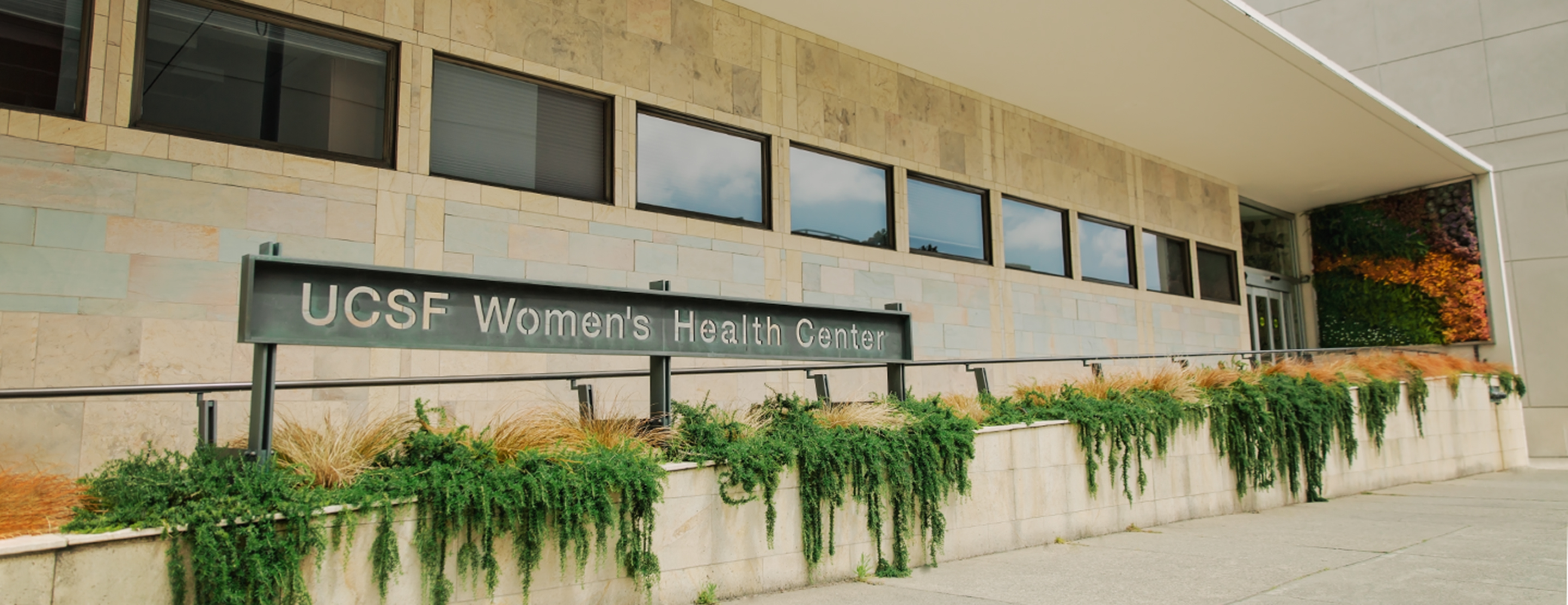
Radiology at the Orthopaedic Institute
UCSF Radiology at the Orthopaedic Institute provides musculoskeletal imaging tests, such as MRI and bone mineral density measurements, as well as image-guided procedures for treating osteoarthritis. We also offer the convenience of same-day X-ray services when you come to the institute for your orthopedic appointment.
The close collaboration between our orthopedic surgery and diagnostic radiology teams translates to better care for patients. Our dedicated radiologists and technologists have particular expertise from many years of working with orthopedic patients.
Our Services
The innovative techniques and technologies available at the Orthopaedic Institute include:
- High-field, high-resolution MRI operating at 3 Tesla used for joint and spine disorders, bone and soft tissue disorders, and rheumatologic diseases. High-resolution MRI at 3 Tesla (the highest-strength magnetic field available for medical practice) allows better detection of abnormalities than standard MRI.
- MR arthrography for better evaluation of shoulder, wrist, elbow and hip joints to find the cause of pain and limited function. This technique (in which we inject the joint with a material that enhances imaging) improves analysis of cartilage.
- MRI techniques to analyze cartilage quality before cartilage is lost, which allows us to assess the risk for osteoarthritis.
- Metal-suppression techniques that allow us to use MRI with patients who have joint replacements, implants and other hardware.
- MR technologies to suppress motion in patients with pain or claustrophobia, allowing us to obtain better-quality images.
- Joint injection for osteoarthritis pain treatment including local anesthetics, corticosteroids and viscosupplements such as hyaluronate and Synvisc (materials similar to joints' natural lubrication fluid).
- Bone mineral density measurements using dual energy X-ray absorptiometry.
- Tomosynthesis (a technique that allows precise evaluation without increased radiation) for better detection of fractures and fracture healing.
MRI and Claustrophobia
While open scanners are helpful for patients with claustrophobia, their image quality is generally worse than that produced by our 3 Tesla scanner. Poor image quality may limit what we can learn from the images.
Keep in mind that:
- Our MRI is shorter, wider and better lit as compared to other MRI machines.
- Our staff understands claustrophobia.
- Imaging of the lower extremities – foot, knee and leg exams – do not require entering the tube completely. Only the leg is in the tube.
- Imaging of the shoulders or spine requires entering the tube, and sedation should be considered. After receiving a sedative, you should not drive a car or operate machinery.
- Our motion-correction imaging allows for shorter exam times and less time spent in the scanner.
Images and reports
Getting your results
A radiologist will review your images and write a report. When finalized, the report and your images will be available in MyChart, UCSF's secure online portal for patients. You can access this report as soon as this occurs, so you may see it before your referring doctor does. Learn more about receiving test results and medical reports in MyChart. If you have questions about a report, please contact your referring doctor.
To get a CD of your medical images and reports, send a request to the UCSF Imaging Library.
Sending images to UCSF
If you need to send medical images that were taken at another institution to your UCSF radiologist, you can do so through our secure online portal. For instructions, visit How to Send Radiology Images to UCSF.
Doctor referral required
Our locations (1)
Our team
How to send radiology images to UCSF
Before your appointment, securely upload images for our radiologists to review.
Accessing your test results in MyChart
Find out how to access your test results and medical reports.
Clinical trials
Focused Ultrasound to Promote Immune Responses for Undifferentiated Pleomorphic Sarcoma
Proportion of participants with device-related adverse events will be determined by evaluating the incidence and severity of any device-related complications from the treatment day visit through the time of surgery or last post-tr...
Recruiting
Support services
Plan your visit
What to Bring
- Photo I.D.
- Health insurance card
- List of your medications, including dosages
- Written request form for the procedure, if your doctor gave you one
- List of questions you may have
- Device or paper for taking notes
Related clinics
Our research initiatives
-
UCSF Department of Radiology and Biomedical Imaging Research
The UCSF Department of Radiology and Biomedical Imaging is home to many state-of-the-art research labs, all working to use imaging technologies to improve the understanding, diagnosis and treatment of disease.

