Mammogram
A mammogram is a low-dose X-ray image of the breast. It can reveal small abnormalities in this tissue long before patients see or feel a change.
Mammograms are used for two main purposes: screening and diagnosis. A screening mammogram is a routine test used to detect breast cancer at its earliest stages, when it’s easiest to treat. A diagnostic mammogram is a more in-depth test to investigate suspicious symptoms (such as a lump) or findings from a previous test.
Different health organizations have different guidelines for when to begin mammogram screenings and how often to get them. Many women start annual screenings at age 40, but some patients with a higher-than-average breast cancer risk may benefit from earlier or more intensive testing.
When examining your mammogram images, the radiologist will look for:
- Calcifications – These are tiny calcium deposits in breast tissue that look like white spots on the X-ray image. Certain calcification patterns can indicate breast cancer or precancerous changes. If you have an abnormal calcification pattern, your doctor may recommend a breast biopsy.
- Masses – Lumps, which doctors call masses, may be cancer but more often are caused by benign (noncancerous) breast conditions. Common benign lumps include cysts (fluid-filled sacs) and fibroadenomas (solid lumps).
Most clinics now use digital mammography rather than film. Digital mammography allows your doctor to view your images on a computer screen from multiple angles.
Digital breast tomosynthesis (DBT, also known as 3D mammography) is an advanced type of digital mammography. With DBT, we can take multiple images of a breast from different angles so the radiologist can see through overlapping tissue.
DBT is better than conventional mammography at early detection of breast cancer. DBT also produces more detailed images, resulting in fewer inconclusive mammograms that require patients to come back for additional testing.
How to prepare
To identify changes over time, your radiologist may want to compare your latest mammogram images with your past images. Learn how to send images taken at other institutions to your radiologist at UCSF Health.
On the day of your mammogram, don’t use deodorant, perfume, lotion or powder on your breasts or under your arms. These products can interfere with the imaging process.
If you are pregnant or breastfeeding or have ever had a breast biopsy, make sure your doctor and mammogram technologist are aware of these things. Screening mammograms aren’t performed during pregnancy or when breastfeeding.
What to expect
When you arrive, you’ll be asked to undress from the waist up, change into a gown, and remove any jewelry from your neck and chest area.
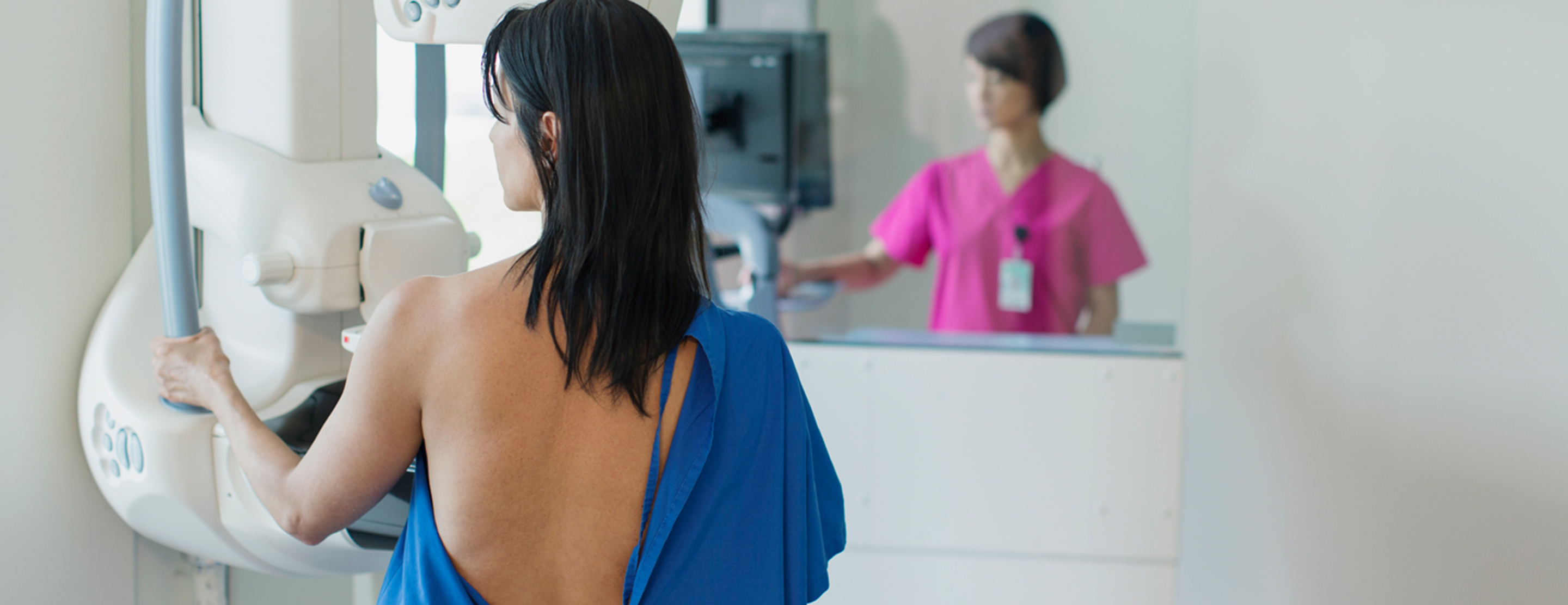
Depending on the equipment being used, you either sit or stand while the images are taken. The technologist places one of your breasts on the X-ray plate. A device presses against the breast to flatten it while the image is taken. This may feel cold and uncomfortable for several seconds.
The technologist takes images from several angles and may ask you to hold your breath while each image is taken. These steps are repeated for your other breast.
The entire mammogram process usually takes about 20 minutes.
Getting the results
If your breast tissue shows no signs of masses or calcifications, your mammogram is considered normal.
If your screening mammogram reveals breast tissue changes, your doctor will want to investigate further (see "Follow-up testing," below). Most of the time, these abnormalities turn out to be benign (not cancerous). You may also be asked to come back for additional images if the radiologist couldn’t clearly see any part of your breasts on the initial mammogram.
Follow-up testing
Your doctor may order the following tests to investigate suspicious findings on a mammogram:
- Additional mammogram views, including magnified images of a specific area
- Ultrasound of the breast
- MRI exam of the breast
If a mammogram or ultrasound reveals abnormalities, we may need to perform a biopsy to collect a small sample from the area of concern. A pathologist (a doctor who specializes in tissue analysis) will look for cancer cells in the sample. A biopsy is the only way to confirm a breast cancer diagnosis.
Types of biopsies include:
Risks
The radiation dose from a mammogram is small and doesn't significantly affect your cancer risk.
Screening mammograms aren't done when a patient is pregnant or breastfeeding. If you need a diagnostic mammogram while pregnant, the technologist will cover your abdomen with a lead apron.
Mammography myths
| Myth 1: Mammogram radiation increases my chance of getting cancer. | Fact: Modern mammography equipment uses a very small dose of radiation for each X-ray. The benefits of this procedure far outweigh the risk from this radiation dose, which doesn't meaningfully affect your cancer risk. |
| Myth 2: Mammograms don't work on younger women. | Fact: Young women tend to have denser breast tissue than older women, which can make abnormalities harder to find using standard mammography equipment. However, 3D mammography produces higher-quality images that helps doctors detect abnormalities in dense breasts. |
| Myth 3: It doesn't matter where I have my mammogram. | Fact: Studies show that radiologists who specialize in mammography, as well as those who interpret a large number of mammograms, are better at detecting cancer early. We recommend imaging centers that specialize in mammography, use the latest equipment, and are designated a breast imaging center of excellence by the American College of Radiology. If possible, use the same center each year so your radiologist can easily compare your latest mammogram to your past images. |
UCSF Health medical specialists have reviewed this information. It is for educational purposes only and is not intended to replace the advice of your doctor or other health care provider. We encourage you to discuss any questions or concerns you may have with your provider.
Review Date: 12/10/2022
The information provided herein should not be used during any medical emergency or for the diagnosis or treatment of any medical condition. A licensed physician should be consulted for diagnosis and treatment of any and all medical conditions. Call 911 for all medical emergencies. Links to other sites are provided for information only -- they do not constitute endorsements of those other sites. Copyright ©2019 A.D.A.M., Inc., as modified by University of California San Francisco. Any duplication or distribution of the information contained herein is strictly prohibited.
Information developed by A.D.A.M., Inc. regarding tests and test results may not directly correspond with information provided by UCSF Health. Please discuss with your doctor any questions or concerns you may have.


