


Diagnosis
Neurology & Neurosurgery
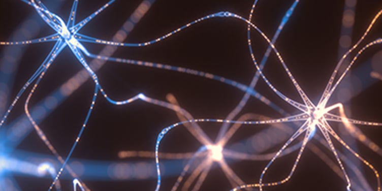
Dural Arteriovenous Fistulae (DAVF)
Dural Arteriovenous Fistulae (DAVF)
Diagnosis
The following tests may be used to diagnose your dural arteriovenous fistulae (DAVF) and help identify its size, location and blood-flow pattern.
- Cerebral Angiography This X-ray exam shows the structure of blood vessels and is the most important test in diagnosing DAVF. A harmless dye, visible on X-rays, is injected into an artery that supplies blood to the brain. As the dye flows through blood vessels to the brain, it will show any obstructions or leaks.
- Computed Tomography (CT) Scan During this test, X-ray beams are used to create a 3-D image of the brain and may help identify any bleeding or hemorrhage. For more precise images of blood vessels, the cerebral angiography and magnetic resonance angiography (MRA) are performed.
- Magnetic Resonance Angiography (MRA) An MRA uses magnetic resonance imaging to create detailed images of blood vessels. Using a strong magnetic field, it generates a 3-D image of the brain to detect, diagnose and aid the treatment of DAVFs and other vascular disorders. In some cases, a dye is injected intravenously.
UCSF Health medical specialists have reviewed this information. It is for educational purposes only and is not intended to replace the advice of your doctor or other health care provider. We encourage you to discuss any questions or concerns you may have with your provider.