Arteriovenous Malformation

Overview
Arteriovenous malformations are abnormal tangles of arteries and veins that belong to a group of disorders known as vascular malformations. Although not completely understood, they typically develop in the womb or soon after birth and may be linked to genetic mutations. When AVMs are located in the brain or spinal cord, they're called neurological AVMs.
Although people are born with AVMs, symptoms may not develop until adulthood, often between 20 to 40 years of age, after the condition progresses, and in most adults, they cause no health problems. About 1 percent of those affected, however, experience life-threatening complications.
Serious complications occur when AVMs:
- Disrupt the flow of blood and reduce the amount of oxygen reaching the brain or spine
- Rupture and bleed into surrounding tissues
- Become so large that they compress or displace parts of the brain or spinal cord
The most severe risk is bleeding, called a hemorrhage, in the brain, which can lead to a debilitating or fatal stroke.
Our Approach to Arteriovenous Malformation
Our team of neurologists, neurosurgeons and neuroradiologists treats more than 100 people with arteriovenous malformations (AVMs) each year. We offer the full range of treatments, including embolization to block blood flow to the AVM before surgical removal, as well as noninvasive radiosurgery, which uses a precisely targeted, high dose of radiation to destroy the blood vessels feeding the AVM.
Awards & recognition
-

Among the top hospitals in the nation
-

Best in California and No. 2 in the nation for neurology & neurosurgery
Signs & symptoms
Most people with arteriovenous malformations don't experience any symptoms. For those that do have symptoms, the most common include:
- Abnormal sensations such as numbness or tingling
- Dizziness
- Headache
- Seizures
Symptoms can vary widely, depending on the location of the AVM. Other symptoms are memory loss, muscle weakness, and visual distrubances, such as partial vision. Some researchers believe the condition can cause subtle learning or behavioral disorders, long before other symptoms emerge.
The most serious complication is bleeding in the brain, resulting in a stroke.
Damage from AVMs tend to build-up over time. In women, pregnancy can sometimes trigger symptoms due to increases in blood volume and blood pressure.
If no symptoms occur by the time people reach their late forties or early fifties, AVMs typically remain stable.
Because AVMs may not produce symptoms, they may be discovered during treatment for other disorders.
In children, AVMs are the leading cause of hemorrhagic stroke.
Diagnosis
The following tests may be used to diagnose your arteriovenous malformation (AVM), as well as help identify its size, location and blood-flow pattern.
- Angiography This special X-ray exam shows the structure of a person's blood vessels and is essential in the diagnosis and treatment planning of AVMs. During this procedure, a harmless dye that can be seen on X-rays is injected into an artery that supplies blood to the brain. The dye follows the path of the brain's blood flow and can show any obstructions or leaks.
- Computed Tomography (CT) Scan With this test, X-ray beams are used to create a 3-dimensional image of the brain. A CT scan typically can detect bleeding into the brain, called a hemorrhage, which indicates an AVM.
- Magnetic Resonance Angiography (MRA) This procedure is a magnetic resonance imaging (MRI) study of the blood vessels and provides detailed images of blood vessels. Using a strong magnetic field, an MRI generates a 3-D image of the brain to detect, diagnose and aid the treatment of vascular disorders. The procedure is painless.
Treatments
Today, there are many safe and highly effective therapies available to treat arteriovenous malformations (AVM). These include surgery, radiation therapy, embolization and radiosurgery using a device called a Gamma Knife.
- Conventional Surgery In many cases, surgery may be recommended to completely remove the AVM. This is appropriate when the AVM is small and located on the surface of the brain or spinal cord. When the AVM is deep in the brain, other minimally invasive techniques are used to prevent damage to surrounding tissue.
- Embolization Embolization is a technique used to reduce blood flow to the AVM by obstructing surrounding blood vessels. During this procedure, the AVM is filled with specially designed coils, glues or spheres that plug its vessels and decrease the flow of blood. Embolization usually doesn't permanently resolve the AVM but makes it more manageable for future procedures such as surgery.
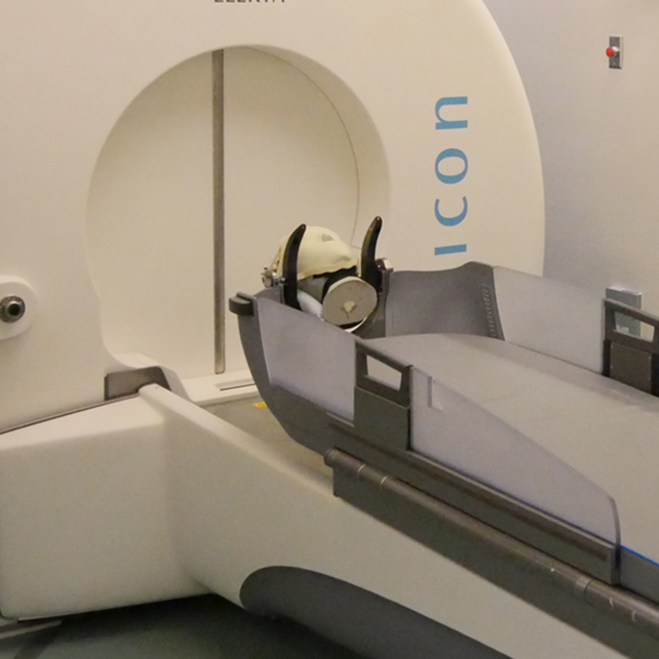
- Radiosurgery The Gamma Knife, an advanced radiosurgery treatment, is often recommended for people with complex, deep-seated or brain-stem AVMs. Despite its name, the Gamma Knife isn't a knife at all. It delivers a single, very finely focused, high dose of radiation precisely to its target, while causing little or no damage to surrounding tissue. The high dose of radiation damages and eventually closes the walls of the blood vessel. Radiosurgery can be used alone or in combination with other treatments, such as conventional surgery.
UCSF Health medical specialists have reviewed this information. It is for educational purposes only and is not intended to replace the advice of your doctor or other health care provider. We encourage you to discuss any questions or concerns you may have with your provider.


